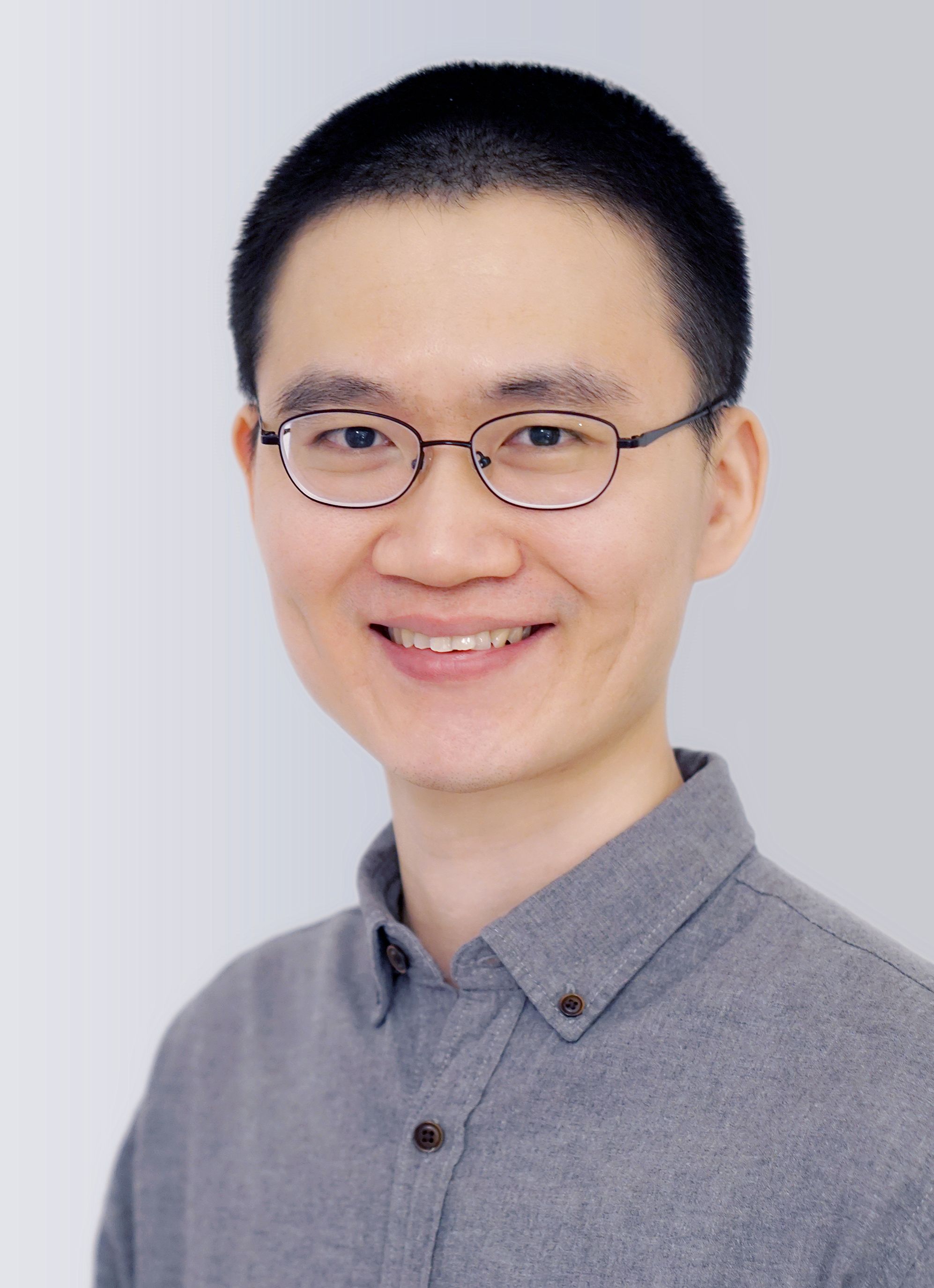
| E-mail: chengshiya@126.com |
Biography
2024-Present Professor, TaiKang Medical School (School of Basic Medical Sciences), Wuhan University
2024-Present PI, TaiKang Center for Life and Medical Sciences, Wuhan University
2016-2024 Postdoc, Max Planck Institute for Multidisciplinary Sciences
2014-2016 Postdoc, National Institute of Biological Sciences, Beijing
2009-2014 Ph.D., Peking Union Medical College / National Institute of Biological Sciences, Beijing
2005-2009 B.S., Sichuan University
Research
Impaired oogenesis is one of the major causes of infertility. Oocytes accumulate a large number of maternal components, including mRNAs, proteins and different types of organelles, to support oocyte maturation and early embryonic development. Our research has been focused on the accumulation and regulation of these maternal components. We combine state-of-the-art imaging, genetics, biochemistry, molecular biology and multi-omics techniques to investigate the roles of these factors in oocyte maturation and early embryonic development, to elucidate the pathogenic mechanisms of infertility caused by abnormalities in these factors, and to explore potential therapeutic approaches.
Representative Publications
1. Cheng, S. and Schuh, M. (2024). Translational control of prophase I arrest in mouse oocytes. Nat Commun. (Under revision)
2. Cheng, S.#, Altmeppen, G.#, So, C., Welp, L.M., Penir, S., Ruhwedel, T., Menelaou, K., Harasimov, K., Stützer, A., Blayney, M., Elder, K., Möbius, W., Urlaub, H., and Schuh, M. (2022). Mammalian oocytes store mRNAs in a mitochondria-associated membraneless compartment. Science 378, eabq4835.
3. Zaffagnini, G., Cheng, S., Salzer, M.C., Pernaute, B., Duran, J.M., Irimia, M., Schuh, M., Böke, E. (2024). Mouse oocytes sequester aggregated proteins in degradative super-organelles. Cell 187, 1109-1126.e21.
4. Fu, L., Weiskopf, E.N., Akkermans, O., Swanson, N.A., Cheng, S., Schwartz, T.U., and Gorlich, D. (2024). HIV-1 capsids enter the FG phase of nuclear pores like a transport receptor. Nature 626, 843-851.
5. Harasimov, K., Gorry, R.L., Welp, L.M., Penir, S., Horokhovskyi, Y., Cheng, S., Takaoka, K., Stützer, A., Frombach, A., Taylor-Tavares, A.L., Raabe, M., Haag, S., Saha, D., Grewe, K., Schipper, V., Rizzoli, S.O., Urlaub, H., Liepe, J., Schuh, M. (2024). The maintenance of oocytes in the mammalian ovary involves extreme protein longevity. Nat Cell Biol.
6. So, C.#, Cheng, S.#, and Schuh, M. (2021). Phase Separation during Germline Development. Trends Cell Biol 31, 254-268.
7. Hu, J.#, Cheng, S.#, Wang, H., Li, X., Liu, S., Wu, M., Liu, Y., and Wang, X. (2019). Distinct roles of two myosins in C. elegans spermatid differentiation. PLoS Biol 17, e3000211.
8. Cheng, S., Liu, K., Yang, C., and Wang, X. (2017). Dissecting Phagocytic Removal of Apoptotic Cells in Caenorhabditis elegans. Methods Mol Biol 1519, 265-284.
9. Cheng, S., Wang, K., Zou, W., Miao, R., Huang, Y., Wang, H., and Wang, X. (2015). PtdIns(4,5)P(2) and PtdIns3P coordinate to regulate phagosomal sealing for apoptotic cell clearance. J Cell Biol 210, 485-502.
10. Wu, Y., Cheng, S., Zhao, H., Zou, W., Yoshina, S., Mitani, S., Zhang, H., and Wang, X. (2014). PI3P phosphatase activity is required for autophagosome maturation and autolysosome formation. Embo Rep 15, 973-981.
11. Cheng, S., Wu, Y., Lu, Q., Yan, J., Zhang, H., and Wang, X. (2013). Autophagy genes coordinate with the class II PI/PtdIns 3-kinase PIKI-1 to regulate apoptotic cell clearance in C. elegans. Autophagy 9, 2022-2032.
12. Wang, H., Lu, Q., Cheng, S., Wang, X., and Zhang, H. (2013). Autophagy activity contributes to programmed cell death in Caenorhabditis elegans. Autophagy 9, 1975-1982.
13. Li, W., Zou, W., Yang, Y., Chai, Y., Chen, B., Cheng, S., Tian, D., Wang, X., Vale, R.D., and Ou, G. (2012). Autophagy genes function sequentially to promote apoptotic cell corpse degradation in the engulfing cell. J Cell Biol 197, 27-35.